CHOOSE YOUR CLINIC
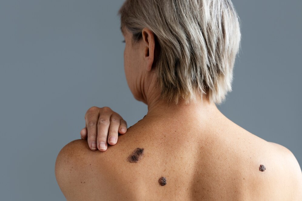
Mole mapping is an advanced dermatological examination that uses high-resolution photography to document and monitor moles and skin lesions across the entire body over time.
Mole mapping helps detect early changes in moles that may indicate melanoma or other forms of skin cancer, often before symptoms become visible.
Mole mapping is recommended for individuals with multiple moles, atypical moles, a personal or family history of skin cancer, or significant sun exposure.
The procedure involves taking standardized, full-body photographs that are securely stored and compared during follow-up visits to identify any new or changing lesions.
Yes. Mole mapping is a non-invasive, painless examination that does not involve radiation or skin damage.
The frequency depends on individual risk factors, but most patients benefit from annual or dermatologist-recommended follow-up exams.
Yes. By accurately tracking mole changes over time, mole mapping helps dermatologists distinguish benign lesions from suspicious ones, reducing unnecessary procedures.